Main Hall - Show Case B - Diagnosis
1 - The Study of the Blood - Measuring Blood Pressure
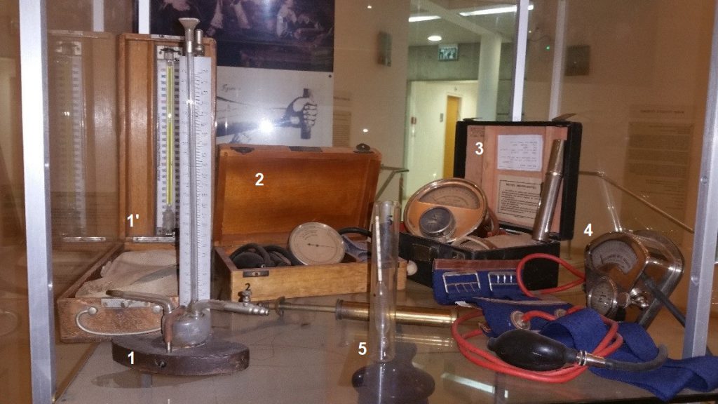
- Scipione Riva Rocci’s sphygmometer, 1895
In 1881, Samuel Sigfrid von Basch developed the first, rather inaccurate sphygmomanometer (from the Greek sphygmus, pulse) to measure blood pressure from outside the body based on an open ended mercury column.
Since the blood pressure within an artery is equal to the force necessary to collapse the vessel, it seemed reasonable that this force might be measured. The sphygmometer supplied the force as well as the manometer reading that corresponds to the pressure when the pulse bellow it ceased. To be effective, the artery must be pressed against an underlying bone.
Fifteen years later, Riva Rocci, an Italian physician produced a model with an inflatable arm band which is virtually the same as that used today. The first practical clinical blood pressure measuring instrument based on an open ended single, straight mercury column. This instrument made blood pressure measurement practical. This simple but effective design continued for years (i.e. Becton-Dickson “pocket” manometer, c. 1926 (SEE CASE NO. 3 AT THE 3ED. FLOOR) that is the direct descendent of the Riva Rocci’s sphygmometer
1’. A similar sphygmomanometer in a wooden case c. 1930
- Baumanometer, c. 1917. Desk model in its wooden box.
- Clock type sphygmometer based on the Bourdon hollow curved springy tube that changes its form when air or liquid was pumped in, c. 1920 (Dr. Naphtali Weise)[1]. To the left: Brass hand pump used in some of the sphygmometers, c. 1915
- Aneroid sphygmometer, c. 1909
This instrument is based on well-known principle of the aneroid (dry) barometer.
- Specific-weight measuring glass tube used to measure the specific weight of urine. The weighted tube sinks according to the specific weight of the liquid where it is immersed.
This specimen was made c. 1935
[1] Dr. Naphtali Weise born 1868 in Odesa, came to Israel 1897 worked in Rosh-Pina, Zichron Yakov and Jerusalem where he died 1935.
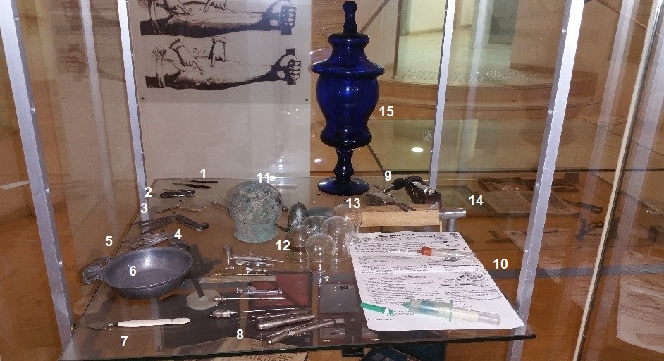
2 - From Leeches to Letting of Blood, Transfusion and Infusion
Until the modern era little was known of the true causes of most diseases and many treatments modalities were based on the assumption that if the “ailing factor” that causes the illness will be removed the patient will be healed. Blaming the “bad blood”, bloodletting was used for most diseases and ailments. It has been used even to treat anemia, malnutrition, Tuberculosis and Malaria and in many cases it harmed more than helped. Patients were bled with leeches, by cuts and incisions in various parts of their body, some aiming to the veins or even arteries. Sometimes, a multitude of incisions (“scarification”) were made in order to increase its efficacy.
“Cupping”– vacuum under a small glass that pulls the “bad blood” away could help the bleeding. The combination of cupping and bleeding incisions was termed a “wet cupping” and when the cupping glass was applied over an intact skin, forming a subdermal or intradermal hematoma it was called “dry cupping”.
Blood Letting Instruments
Though any sharp object could be used, the most common ones were sharp flint tools, arrows and arrows heads, cutting and stabbing tools and eventually, hollow needles.
There was a clear distinction between a knife, surgical knife and lancet that was specifically used for
blood-letting. The lancets were small double-edged sharp blades protected by swiveling plates that also served as handles. The shape of the blade did not change through history (SEE ROMAN ACALPEL IN CASES No. C & I IN THE GALLERY). The blood was drained into “bleeding bowls” made of porcelain, pewter, silver or gold.
- Two classical Lancet used for bloodletting and incisions, c.1800 Turtle shell guard and steel blade
- Two small lancets, c. 1750. Turtle shell guard and steel blade in a silver case
- “Modern” lancet, c. 1890, stainless steel
4+5. Fleams
Fleams were penknife-looking instruments with several (3-4) different blades, one straight and pointed used for stabbing and the other for lateral incision used mainly for bloodletting in human and veterinary medicine.
An example of an early fleam can be seen in CASE ” I” IN THE GALLERY a bleeding fleam with several sizes of blades, c.1750 Copper & horn handle, steel blades. The owner, John White engraved his name on the handle together with the act of puncturing the skin. The pointed blade he engraved is very similar to the modern surgical blade (no.11) or the pointed straight Roman scalpel.
- An early iron, three blades fleam c. 1700
- Large fleam, c.1850 Horn handle and three steel blades
- Blood-letting bowl, c.1750, pewter
- Large lancet, c. 1800, Ivory handle, steel blade (SEE THE OLDER, ELABORATE JADE HANDLE OF THE LANCET IN CASE “I” IN THE GALLERY)
- Bleeding needles, c. 1900
Some were carried around in a specially made metal cylinders (a) and were designed to allow the pouring blood to flow directly into the bleeding bowl.
Later needles (b, d) were designed to be connected to tubes for transfusion: transferring blood between two humans or between animal and human: Transfusion (see Laurenz Heister textbook, Chirurgie, printed Nürenberg, 1731) (c) Vein obturator for an open phlebotomy, designed to open the collapsed vein to ease the introduction of blunt transfusion tubes (cannulas), 2 gold plated cannulas (d). In some cases the doctor carried a set of 3-4 bleeding-transfusion cannulas with an introduction trocar at its end in a small compact carrying case.
- Bleeding syringe, Park-Davis, 1902. Shaped like a syringe but has a large bore needle with cleaning rod instead of a plunger to clear blood clots.
- The study of the blood
Once the blood was out of the vessel it could be studied. After the invention of the microscope it did not take long to discover the various bodies of the normal blood and the changes in number and shape in pathological conditions. It became important to find cost-effective and simple means to collect blood samples.
Hypodermic syringes and bloodletting needles were the state of the art until they were replaced by the BD (Becton Dickenson USA) “Vacutainer” (10a). It is a 1980-patented vacuum filled test-tube for collecting blood samples.
Strange enough, we were able to find a similar product dated 1928 (10b). The Behring “Venule”, produced in Germany by Bayer 50 years before the USA “Vacutainer” consisted of an external wooden case, an inner metal sheet protective cylinder, a glass vacuum-filled blood collecting tube with an integrated needle and inner valve. The discovery of this exemplar undermined the BD “Vacutainer” patent and market exclusivity.
- Cupping glasses
Cupping was used since Babylonian time to clear away the “bad blood”. Bronze and earth-ware cupping “glasses” were used later by the Egyptians, Greeks and Romans transferring it by the Arabs to the western medicine. Two kinds of cupping were used: “dry” cupping (formation of hematoma) or “wet” cupping, a combination of vacuum cupping and scarification that allows the blood to flow out of the body. From left to right: A. An extremely rare Babylonian bronze cupping “glass”, c.1000 BC, B. A rare Early Arab glass c. 1000 AC, C. a Bedouin tin “cupping glass” ~ 100 years old, D. a Roman c.100 AC suction glass and E. “modern” (50-100 years old) cupping glasses.
- Automatic spring loaded lancets
Four different designs: These could be in the form of small, spring loaded sharp blades that could be used for vaccination (Pirquet’s vaccination lancet, late 19th century) or bloodletting. Being automatic, they were very easy to use even by non-experienced healers. (SEE CASE “I” AT THE GALLERY).
- 12 Blades Scarificator
Spring loaded blades (12!!). Many models were made from the 17th century on for wet cupping (SEE CASE “I” AT THE GALERY).
- Spring loaded “modern” version of the Bauncheidt’s Lebenswecker (life awakener in German), 1851. The original old version with the ebony hollow tube contained a handle with coiled spring attached. When pushed the 30 sharp needles punctured the skin. (SEE CASE “I” AT THE GALLERY). This 19th century version has an adjustable head that controls the depth of the protruding needles. These and similar instruments were used mainly together with cupping. Similar instruments were used at the same time for vaccination.
- Leech jar, 19th century Blue glass: Kept mainly in barber shops where the barbers also offered the services of bloodletting, cupping and even some surgical procedures.
- Leech tube, 18th century
The use of leeches was very common for most ailments. Very often leeches had to be placed on a very specific site (i.e. anus, vagina, or a specific skin area). Not always the leech agreed with its next lunch site and tried to wriggle to more appetizing scenery. The leech tube served to force-apply the leech to its desired pasture. The leech was inserted into the tube headfirst, then directed to the desired spot and left until the leech attached and started to feed. These tubes were rediscovered 20 years ago with the development of microsurgery and the need of placing the leeches on a specific operated area to drain engorged flaps or transplanted organs.
For general questions and questions about accessibility, please email us at info@art&historymuseum.org
3 - Use of the Collected Blood: Study and Transfusion.
Laurenz Heister textbook, Chirurgie, printed Nürenberg, 1731 (facsimile) is one of the most influential illustrated medical textbooks of its time. By then the bleeding techniques were developed to the point that blood vessels could be identified, selectively obstructed by tourniquet and punctured or bled by hollow needle with blood removed or introduced (transfused) at will. Using metal tubes, Heister’s book shows the technique of transfusing blood from human to human or from a calf to human.
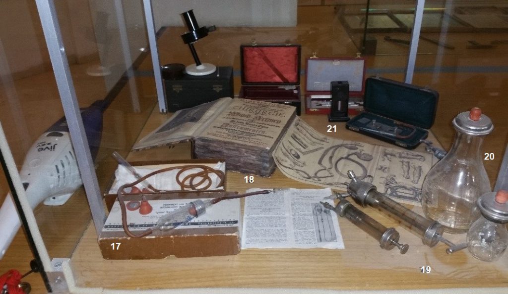
- The first commercial system of infusion: Rubber tubes, glass and metal drop- chamber and filters with an aluminum tourniquet handle, 1935. Such systems were used until the 50’s when rubber, glass and metal was replaced by plastics.
- Two versions of Dieulafoy’s aspirating syringe used also for blood transfusion. Aspiration of blood from the donor in one inlet and with a turn of the syringe head to transfuse it to the host, c. 1900.
- Two infusion bottles, c.1940
- Hematometers
One of many similar systems used since the turn of the 19th century for measuring Hematocrite (the concentration of hemoglobin in the blood) with its small automatic (Pirquet’s type) lancet.
Social Connect
The Rosenberg Museum of Medicine – All Rights Reserved Ben-Gurion University of the Negev – Faculty of Health Sciences