Main Hall - Show Case C (Left side)- Diagnosis
1 - Inspection
With the development of optics and the invention first of the magnifying lens and later the microscope by Leevwenhoek (1668-1783) a new era in science and medicine began. The microscope allowed the discovery of the living cells and the tissue infrastructure and function. It also allowed the discovery of the microorganisms and the pathogenic germs (i.e. bacteria). The second step was the development of small electrical illumination means (end 19th century) for dark recesses (and the body is practically made of such recesses). The third step was developing means to carry light and vision (magnified or not) into these body recess. The first were rigid tubes with a system of lenses and small electrical bulbs. Nowadays we use flexible, very thin (~1 millimeter) optic fibers that allow us to illuminate, see and even perform complicate procedures in most areas of the body through a small incision (including skull, upper and lower respiratory and digestive tracts, abdominal and chest cavities, blood vessels including the heart, joints etc.).
Microscopes
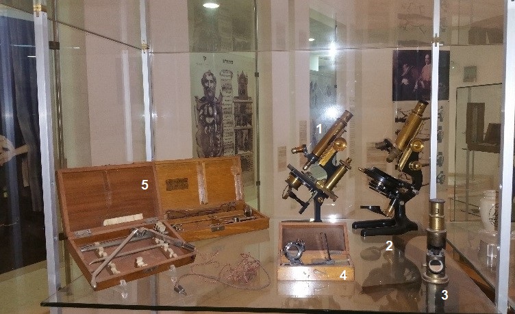
- Watson & son London microscopes, 1890. A typical large microscope
- Zeiss microscope, 1885, Germany. In 1890 Bausck & Lomb produced their version of this microscope in the USA
- A Small continental (British) Martin type microscope, c.1800
- Accessory set of mirrors and filters for the continental-type microscope
- Two Cystoscopes, c. 1930
A rigid tube with a system of lenses and electrical bulb for illumination. This instrument is introduced through the urethra, allowing visualization and interventions of the urinary bladder. Similar instruments were developed and used for visualization of the lungs and upper airways, stomach, etc. Today these instruments are replaced by minimal invasive optic-fiber systems that allow visualizing deep body structures trough thin, flexible, guided and versatile wires.
2 - Auscultation, Percussion and Temperature Measurement.
Auscultation: The art of listening to the body sounds
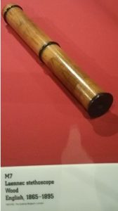
Listing to the body’s sound has been practiced since early times by the physician putting his ear against patient’s chest, back or abdomen. Imitating children transferring voices between two boxes along a string or a tree log, Theophile Hycinte Laennec invented in 1816 the first stethoscope: a wooden tube (23 cm. long and 4 cm in diameter), mainly used for the study of the cardiac and pulmonary sounds. The stethoscope became quickly very popular in most countries, except Republican France, where doctors refused at first to use this important invention because Laennec was a supporter of the monarchy.
Monaural stethoscopes
Elastic, straight-grain and dense woods as ebony and cedar increase the intensity of vibrations. When the stem bulk was reduced by the mid-19th century, the intensity of sound was further increased. Solid wood stethoscopes were used to listen to solid body masses such as the heart or fetal heart. However, surface friction noises competed with internal sounds. By boring a central hole, the transmission of sounds through the air added sounds not otherwise heard. The air column and the bore diameter should be as short and small as possible. Made of most solid materials, stethoscopes became (and still are) very much the physician’s trademark. Some could be dismantled (“composition” monaural stethoscope) and the chest piece attached to the stem at its side transforming it to a percussion hammer allowing the doctor also to stick it on their hat (not unlike a feather). Present day doctors carry their stethoscopes as a status symbol not on their hats but in plain sight on their neck…
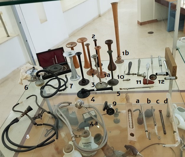
1-5. Early monaural stethoscopes
- Goblet shape interior, c.1840 stethoscope
- 1a, b, two sizes Pinard’s wooden fetal heart’s sound stethoscopes, c 1900-20 and very long one ( Boris Pliskin, Tel Aviv) * that allows some distance from the patient
- Fruitwood, Grumbridge, c.1840 stethoscope
- Fruitwood composition monaural stethoscope, c. 1850
- Composition Monaural stethoscopes, c.1900, vulcanized rubber (“Bakelite”)
4a. A similar Monaural stethoscope, c.1900, promoting “Salicylopyrin Motopyrin Phennin: Motor”
- 2 Composition Monaural Hawksley’s metal and rare aluminum stethoscopes, c. 1900,
Binaural stethoscopes
The use of both ears for listing eliminated interfering noises and increased the amount of sound reaching the listener. At first the tubing was made of India Rubber. Today it is of latex or silicone rubber. It should be thick, about 25 cm long. The internal caliber, including the metal ear tubes and the central hole of the chest piece should be of 3-4mm.
6-7. 3 flexible compact phonendoscopes, c. 1894 the “new “ one has been patented 1897
Bazzi and Bianchi’s 2 smaller, detachable localizers and wounding tubing with vulcanite ear pieces. This new concept used only a column of air as the conducting medium. Chest sounds were loud and clear and without much interference by any positional change from the patient or his physician. It contained a removable metal rod (localizer) that could be attached to the membrane in order to listen to a very small and well defined area.
- An early Cammann 1855/Palmer’s binaural stethoscope, c. 1880
A circular spring replaced the elastic band used also as a caliper for measuring the chest, head, etc.
8a A modified Sprague-Bowls / Meredith twin reversible chest piece 1926-8
- An “Art deco” metal binaural stethoscope, c. 1930
Percussion
Egyptians and Greeks used percussion to investigate solid organs (liver) or cavities (abdomen and chest). Special percussion devices were developed during the 17-18 century
- 2 Killiard percussor c. 1860 ebony, and mother-of-pearl handles and rubber percussion tips
- Tomahawk and Taylor’s 2 percussors/hummers, c. 1910, pattern with rubber edge
- Neurological hammer, c. 1950, chrome with small and large heads a. Retractable needle & brush
accurate and fine sensation diagnosis, c 1930
Thermometers
Different types of thermometers from right to left:
a. Clinical thermometers designed for underarm use, c.1800
b. Encased clinical thermometer for oral use, c. 1860
c. Double use (oral and rectal) thermometers in their cases, c.1900
d. Bath thermometers in wooden frame (will float on water) c.1930
3 - Pollution: Breathing Polluted Air: The Cost of Progress
The industrial revolution changed the living environment. Cities became huge, polluted infernos with undrinkable water and unbreathable air. The smoking chimney and the smog (a wet mixture of sooth and noxious fumes) became the hallmark of the “modern city”.

- Three Protective masks, c. 1870
Masks containing vinegar or other “medicaments” impregnated filters (cotton or wool) were worn in the open in order to achieve some protection. They were not very effective and diseases of the respiratory system were very common. Some of these masks were quite lush and quite expensive (1 shilling) see mask 3* with red velvet)
- Two small menthol holders, c.1880
Menthol holders together with perfumed handkerchiefs were used to counter the stink.
- Four Menthol holders (modern version)
18-21. Humidifiers and bronchial inhalers
Upper respiratory tract and lung diseases, some combined with tuberculosis were very common and inspired a plethora of ingenious humidifiers and inhalers designed to alleviate some of the symptoms. The technique of mixing drugs such as saturated “Tinctura of Sramonium” (Thorn-apple leaves), Iodine, and Conium etc. with vapor for inhaling is not new and was used by Natives in many civilizations.
18, 19. 4 different inhalers late 19th early 20th century offering means of insufflating medicinal and natural preparations
- Inhaler, early 19th century, Africa
- Two rare “Dr. Nelson’s” & “Boot’s” improved Inhalers, late 19th early 20th century, Britain
Social Connect
The Rosenberg Museum of Medicine – All Rights Reserved Ben-Gurion University of the Negev – Faculty of Health Sciences